

TISSUE LEVEL OF ORGANIZATION - Human Anatomy and Physiology B. Pharma 1st Semester
Human Anatomy and Physiology
Introduction
• Human Anatomy:Deals with the structure of the human body and the relationship among the structures
• Physiology:Deals with body functions
Scope
Helps us to learn about the structure and functions of human body and the inter relationship
Parameters of normal body health and factors affecting normal body processes can be known
Forms the basis for proper diagnosis of a disease and its effective treatment
Anatomical and Physiological concepts help in efficient surgeries and understanding the pathology of diseases
Basis for advanced scientific studies and serves as a gateway to get into health related careers
Helps in successful maintenance of community and individual health
Subspecialties of Anatomy
Embryology •First 8 weeks of development
Developmental Biology • From fertilization of an egg to death
Cell Biology •Cellular structure and functions
Histology •Microscopic structure of tissues
Surface anatomy •Surface markings of the body
Gross anatomy • Structures – Without the use of microscope
Systemic anatomy •Structures – Specific systems
Regional anatomy •Specific regions of the body
Radiographic anatomy • Structures – Visualized with X – rays
Pathological anatomy • Structural changes with disease
Neurophysiology •Functional properties of nerve cells
Endocrinology •Hormones and their control
Cardiovascular Physiology • Functions of heart and blood vessels
Immunology •Defense against disease causing organisms
Respiratory Physiology • Functions of air passageways and lungs
Renal Physiology •Functions of kidneys
Exercise Physiology •Muscular activities – Changes
Pathophysiology •Functional changes - Disease and aging
Levels of Organization
Living organisms are made up of four levels of organization: cells, tissues, organs, and organ systems. Order the levels of organization for living organisms
Key Points
Cells are the most basic unit of life at the smallest level of organization.
Cells can be prokaryotic (without nucleus) or eukaroyotic (with nucleus).
The four categories of tissues are connective, muscles, epithelial, and nervous tissues.
Organs are made of different types of tissues and perform complex functions. They can be hollow or solid.
Organ systems are groups of organs that perform similar functions or perform functions together.
Many physiological functions are carried out by multiple organ systems working in tandem.
Key Terms
Cell: The smallest unit of life capable of independent reproduction. Generally contains nucleic acid, cytoplasm, a cell membrane, and many other proteins and structures.
Organ: A structure made of different tissues that work together to perform physiological functions.
Organ system: A group of organs and tissues that work together to perform specific functions.
Tissues: A group of similar cells with the same origin that work together to perform the same function.
EXAMPLES
Using the circulatory system as an example, a cell in this system is a red blood cell, the heart’s cardiac muscle is a tissue, an organ is the heart itself, and the organ system is the circulatory system.
An organism is made up of four levels of organization: cells, tissues, organs, and organ systems. These levels reduce complex anatomical structures into groups; this organization makes the components easier to understand.
Level 1: Cells
The first and most basic level of organization is the cellular level. A cell is the basic unit of life and the smallest unit capable of reproduction. While cells vary greatly in their structure and function based on the type of organism, all cells have a few things in common. Cells are made up of organic molecules, contain nucleic acids (such as DNA and RNA), are filled with fluid called cytoplasm, and have a membrane made out of lipids. Cells also contain many structures within the cytoplasm called organelles, which perform various cellular functions.
Cells may be prokaryotic (without a nucleus) in bacteria and archaea (single-celled organisms), or eukaryotic (with nucleus-enclosing DNA) in plants, animals, protists, and fungi. In humans, most cells combine to form tissues, but some cells are found independent of solid tissues and have their own functions. A red blood cell found circulating in the bloodstream carrying oxygen throughout the human body is an example of an independent cell.
Level 2: Tissues
Tissues are a group of similar cells of the same origin that carry out a specific function together. Humans have four different types of basic tissues. Connective tissues such as bone tissue are made up of fibrous cells and give shape and structure to organs. Muscle tissue is made up of cells that can contract together and allow animals to move. Epithelial tissues make up the outer layers of organs, such as the skin or the outer layer of the stomach. Nervous tissue is made of specialized cells that transmit information through electrochemical impulses, such as the tissue of nerves, the spinal cord, and the brain.
Level 3: Organs
An organ is a structure made up of different tissues that perform specific bodily functions. Most organs contain tissues such as parenchyma (used to perform the organ functions), stroma (connective tissue specific to organs) and epithelial. Organs may be solid or hollow, and vary considerably in size and complexity. The heart, lungs, and brain are all examples of organs.
Level 4: Organ Systems
An organ system is a collection of organs that that work together to perform a similar function. There are eleven different organ systems in the human body, each with its own specific functions. One example is digestive system, which is made up of many organs that work together to digest and absorb nutrients from food. While most organ systems control a few specific physiological processes, some processes are more complex and require multiple organ systems to work together. For example, blood pressure is controlled by a combination of the renal system (kidneys), the circulatory system, and the nervous system.
Levels of Organization in Animals: An organism contains organ systems made up of organs that consist of tissues, which are in turn made up of cells.


5. Different Basic Terminologies Used in Anatomy & Physiology


Regional Terms:-


Regional directional terms include anterior and posterior, dorsal and ventral, and lateral and medial.
Learning Objectives
Describe how axes give direction, detail, and location when describing a region of the body
Key Points
Regional terms describe the different parts of the body by the structures and functions of a specific region. The most basic regional terms are the axial and appendicular regions.
Axes use directional terms to describe the location and orientation of a specific region.
The directional term lateral is used to describe structures divided by a left-to-right axis.
Key Terms
ventral: On the front side of the human body or the corresponding surface of an animal, usually the lower surface.
posterior: Nearer the caudal end of the body in quadrupeds or the dorsal end in bipeds.
axis: A line between two points that is used to give direction to an anatomical region.
Regional Terms in Anatomy
Regional terms describe anatomy by dividing the parts of the body into different regions that contain structures that are involved in similar functions. Two primary terms are used to describe the main regions of the body:
The Axial Region makes up the main axis of the human body and includes the head, neck, chest, and trunk.
The Appendicular Region makes up the parts of the human body that connect to the axial region. This includes the limbs and appendages.
These are the two basic categories of regional terms; however, many other terms are used to describe smaller regions within the axial and appendicular regions. For example, the brachial region consists of the arm as a part of the appendicular region, while the abdominal region consists of the abdomen as a smaller part of the axial region.
The abdominal region is subdivided into even smaller regions based on different functions of groups of organs and tissues in that region. If a person is experiencing pain in one part of the abdominal region, then the smaller regional divisions can help determine the organs involved in the problem to better treat symptoms.
Axes Describe Relative Positions
Another method for describing region An axis uses a straight line between two parts of the body to describe a region of the body with linear direction. For example, blood can be said to flow
in a proximal or distal direction through a region marked by that axis. The X, Y, and Z axes of the Cartesian coordinate system are used describe the specific location of an axis in standard anatomical position.
Many types of axes can give regional direction. Any pair of corresponding directional terms can be combined to form an axis (such as proximal-distal for an appendage).
The Dorsoventral axis (DV axis) is formed by the connection of the dorsal and ventral points of a region. The region between the belly (ventral) and back (dorsal) is often described by a DV axis.
The Anterioposterior axis (AP axis) is the axis formed by the connection of the anterior (top) and posterior (bottom) ends of a region. The AP axis of a region is by definition perpendicular to the DV axis and vice-versa.
The Left-to-right axis is the axis connecting the left and right hand sides of a region. It is used to describe the lateral sides of a region, which in humans are often symmetrical around the center of the body. It is perpendicular to both the DV and AP axes.

Different Directional AP Axes in Three Body Segments of a Horse: Axis (A) (in red) shows the AP axis of the tail, (B) shows the AP axis of the neck, and (C) shows the AP axis of the head.
Axes give more clarity and detail for describing the location of an anatomical region. They are commonly used in both zoology and human anatomy, and can be paired with body planes to give even more detail to anatomical direction, region, and location.


When an organism is in its standard anatomical position, positional descriptive terms are used to indicate regions and features
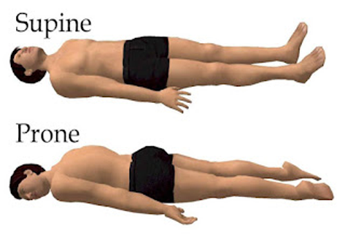
In standard anatomical position, the limbs are placed similarly to the supine position imposed on cadavers during autopsy.
The anatomical position of the skull is the Frankfurt plane. In this position, the lower margins of the orbitals (eye sockets), the lower margin of the orbits, and the upper margins of the ear canals (poria) lie in the same horizontal plane.
Because animals can change orientation with respect to their environments and appendages can change position with respect to the body, positional descriptive terms refer to the organism only in its standard anatomical position to prevent confusion.
Terms
• appendage: A limb of the body.
• supine: Lying on its back, reclined.
• anatomical position: The standard position in which the body is standing with feet together, arms to the side, and head, eyes, and palms facing forward.
The Need for Standardization
Standard anatomical position is the body orientation used when describing an organism’s anatomy. Standardization is necessary to avoid confusion since most organisms can take on many different positions that may change the relative placement of organs. All descriptions refer to the organism in its standard anatomical position, even when the organism’s appendages are in another position. Thus, the standard anatomical position provides a “gold standard” when comparing the anatomy of different members of the same species.
Relative location in the anatomical position: Many terms are used to describe relative location on the body. Cranial refers to features closer to the head, while caudal refers to features closer to the feet. The front of the body is referred to as anterior or ventral, while the back is referred to as posterior or dorsal. Proximal and distal describe relative position on the limbs. Proximal refers to a feature that is closer to the torso, while distal refers to a feature that is closer to the fingers/toes. Medial and lateral refer to position relative to the midline, which is a vertical line drawn through the center of the forehead, down through the belly button to the floor. Medial indicates a feature is closer to this line, while lateral indicates features further from this line.
Standard Anatomical Position in Humans
The standard anatomical position is agreed upon by the international medical community. In this position, a person is standing upright with the lower limbs together or slightly apart, feet flat on the floor and facing forward, upper limbs at the sides with the palms facing forward and thumbs pointing away from the body, and head and eyes directed straight ahead. In addition, the arms are usually placed slightly apart from the body so that the hands do not touch the sides. The positions of the limbs, particularly the arms, have important implications for directional terms in those appendages.
The basis for the standard anatomical position in humans comes from the supine position used for examining human cadavers during autopsies. Dissection of cadavers was one of the primary ways humans learned about anatomy throughout history, which has tremendously influenced the ways by which anatomical knowledge has developed into the scientific field of today.
In humans, the standard anatomical position of the skull is called the Frankfurt plane. In this position, the orbitales (eye sockets), lower margins of the orbits, and the poria (ear canal upper margins) all lie in the same horizontal plane. This orientation represents the position of the skull if the subject were standing upright and looking straight ahead.
It is important to note that all anatomical descriptions are based on the standard anatomical position unless otherwise stated.
Directional Terms:-


Positional terms give precise descriptions of anatomical relationships and allow for consistency when referencing anatomical positions.
Key Points
Descriptions of directional terms include: a) superior (head) and inferior (caudal), b) anterior and posterior, c) lateral and medial, d) deep and superficial, e) proximal and distal, and f) dorsal and ventral.
Directional terms provide comparison of anatomical position by comparing the locations of different structures in the body.
Key Terms
Directional terms: Directional terms are words used to describe the location of an anatomical structure by comparing its position to other structures within the body or within the orientation of the body itself.
Navigating Anatomy with Language
Directional terms provide precise descriptions of a structure’s location. They allow a description of anatomical position by comparing location relative to other structures or within the rest of the body. Standard anatomical terms for direction include:
Superior and inferior (cranial and caudal) are used when referring to parts of the body which are toward an end of the body. Superior structures are toward the head (cranial) while inferior (caudal) structures are toward the feet. Examples include the superior and inferior vena cava, which carry deoxygenated blood away from the head (superior) and from the lower body (inferior) to the heart.
Anterior and posterior are sometimes used in place of superior and inferior, respectively. These words are used more often for animal anatomy and rarely and only with very specific meaning in human anatomy. Anterior refers to the side of the structure facing up in the standard anatomical position while posterior refers to the bottom side. For example, the pituitary gland has an anterior and posterior side, each of which secretes different types of hormones.
Dorsal and ventral are sometimes used in place of anterior and posterior, respectively. Dorsal means the back side or upper side, while ventral means the frontal or lower side. These are mostly used with animal anatomy, but can be used in human anatomy as long as they are describing the side of an appendage. One example is the dorsal fin in fish, found on the upper side of the fish’s body.
Lateral is used to describe anything closer to the sides of the body (toward the arms, in the standard anatomical position), whilemedial is used to describe anything toward the middle of the body. In general, many structures of the human body are bilateral and symmetrical with the middle of the body, such as the lungs or the arms.
Deep refers to structures closer to the interior center of the body. For example, bones in an appendage are located deeper than the muscles. Superficial is used to describe structures that are closer to the exterior surface of the body. For example, the outer layers of skin are superficial to deeper layers of skin.
Proximal and Distal describe one point relative to another. Proximal refers to a point closer to the reference point while distal refers to a point farther away. When describing appendages, the proximal end of the appendage connects the appendage to the body, while the distal end is away from the body.
Body Planes:-

There are three basic reference planes used in anatomy: the sagittal plane, the coronal plane, and the transverse plane.
Key Points
A coronal or frontal plane divides the body into dorsal and ventral (back and front, or posterior and anterior) portions.
A transverse plane, also known as an axial plane or cross-section, divides the body into cranial and caudal (head and tail) portions.
A sagittal plane divides the body into sinister and dexter (left and right) portions.
Body planes have several uses within the anatomy field, including in medical imaging, descriptions of body motion, and embryology.
Key Terms
Coronal plane: Any vertical plane that divides the body into anterior and posterior (belly and back) sections.
Transverse plane: Any plane that divides the body into superior and inferior parts, roughly perpendicular to the spine.
Sagittal plane: Any imaginary plane parallel to the median plane.
What Are Body Planes?
Body planes are hypothetical geometric planes used to divide the body into sections. They are commonly used in both human and zoological anatomy to describe the location or direction of bodily structures. Reference planes are the standard planes used in anatomical terminology and include:
The sagittal plane (lateral or Y-Z plane) divides the body into sinister and dexter (left and right) sides. The midsagittal (median) plane is in the midline through the center of the body, and all other sagittal planes are parallel to it.
The coronal plane (frontal or Y-X plane) divides the body into dorsal and ventral (back and front) portions. It also separates the anterior and posterior portions.
The transverse plane (axial or X-Z plane) divides the body into superior and inferior (head and tail) portions. It is typically a horizontal plane through the center of the body and is parallel to the ground.
While these are the major reference planes of the body, other planes are commonly used in relation to these three. A longitudinal plane is any plane perpendicular to the transverse plane, while parasaggital planes are parallel to the saggital plane.
The coronal plane, the sagittal plane, and the parasaggital planes are examples of longitudinal
Planes.
Anatomical Planes in a Human: There are three basic planes in zoological anatomy: sagittal, coronal, and transverse. A human in the anatomical position, can be described using a coordinate system with the Z-axis going from front to back, the X-axis going from left to right, and the Y-axis going from up to down.
Applications of Body Planes
Medical imaging techniques such as sonography, CT scans, MRI scans, or PET scans are one of the primary applications of body planes. By imaging a patient in standard anatomical position, a radiologist can build an X-Y-Z axis around the patient to apply body planes to the images. The planes can then be used to identify and locate the positions of the patient’s internal organs. Individual organs can also be divided by planes to help identify smaller structures within that organ.
Body planes are used to describe anatomical motion in the X-Y-Z coordinate system that the body moves through. An anatomist could model a limb’s range of motion by measuring which planes the limb can move through and how far it is able to travel.
Anatomical change during embryological development is also described and measured with body planes. For example, during human embryonic development the coronal plane is horizontal, but becomes vertical as the embryo develops into a fetus. In comparative embryology, body planes provide a basis for comparing the ways in which different types of organisms develop anatomically within the womb.
Body Cavities:-

Key Points
The dorsal cavity contains the primary organs of the nervous system, including the brain and spinal cord.
The diaphragm is a sheet of muscle that separates the thoracic cavity from the abdominal cavity.
Special membrane tissues surround the body cavities, such as the meninges of the dorsal cavity and the mesothelium of the ventral cavity.
The mesothelium consists of the pleura of the lungs, the pericardium of the heart, and the peritoneum of the abdominopelvic cavity.
Key Terms
abdominoplevic cavity: The ventral body chamber that contains the abdominal cavity (primarily digestive system) and the pelvic cavity (primarily reproductive system).
dorsal cavity: The cavity in the back of the body that contains the cranial and vertebral cavities, which house the brain and spinal cord respectively.
Thoracic Cavity: The ventral body chamber that contains the pericardial cavity (the heart) and the pleural cavity (the lungs).
By the broadest definition, a body cavity is any fluid-filled space in a multicellular organism. However, the term usually refers to the space where internal organs develop, located between the skin and the outer lining of the gut cavity.”The human body cavity,” normally refers to the ventral body cavity because it is by far the largest one in volume. Blood vessels are not considered cavities but may be held within cavities. Most cavities provide room for the organs to adjust to changes in the organism’s position. They usually contains protective membranes and sometimes bones that protect the organs.

Anatomical terminology for body cavities: Humans have multiple body cavities, including the cranial cavity, the vertebral cavity, the thoracic cavity (containing the pericardial cavity and the pleural cavity), the abdominal cavity, and the pelvic cavity. In mammals, the diaphragm separates the thoracic cavity from the abdominal cavity.
Dorsal
The dorsal cavity is a continuous cavity located on the dorsal side of the body. It houses the organs of the upper central nervous system, including the brain and the spinal cord. The meninges is a multi-layered membrane within the dorsal cavity that envelops and protects the brain and spinal cord.
Cranial
The cranial cavity is the anterior portion of the dorsal cavity consisting of the space inside the skull. This cavity contains the brain, the meninges of the brain, and cerebrospinal fluid.
Vertebral
The vertebral cavity is the posterior portion of the dorsal cavity and contains the structures within the vertebral column. These include the spinal cord, the meninges of the spinal cord, and the fluid-filled spaces between them. This is the most narrow of all body cavities, sometimes described as threadlike.
Ventral
The ventral cavity, the interior space in the front of the body, contains many different organ systems. The organs within the ventral cavity are also called viscera. The ventral cavity has anterior and posterior portions divided by the diaphragm, a sheet of skeletal muscle found beneath the lungs.
Thoracic
The thoracic cavity is the anterior ventral body cavity found within the rib cage in the torso. It houses the primary organs of the cardiovascular and respiratory systems, such as the heart and lungs, but also includes organs from other systems, such as the esophagus and the thymus gland. The thoracic cavity is lined by two types of mesothelium, a type of membrane tissue that lines the ventral cavity: the pleura lining of the lungs, and the pericadium lining of the heart.
Abdominopelvic
The abdominoplevic cavity is the posterior ventral body cavity found beneath the thoracic cavity and diaphragm. It is generally divided into the abdominal and pelvic cavities. The abdominal cavity is not contained within bone and houses many organs of the digestive and renal systems, as well as some organs of the endocrine system, such as the adrenal glands. The pelvic cavity is contained within the pelvis and houses the bladder and reproductive system. The abdominopelvic cavity is lined by a type of mesothelium called the peritoneum.
6. Summary
Anatomy and Physiology: Study of Structure & functions of human body
Levels of organization: Atomic level to organismal level
Each of the body systems has several functions and these are interconnected
Various systems work together to maintain homeostasis
Anatomical terms, body planes and cavities: Help to describe the body structure
Regional and directional terms: Useful to differentiate body landmarks


0 Comments: