

Urinary system
Objectives
At the end of this lecture, student will be able to
• Describe the functions of urinary system
• Describe the external and internal gross anatomical features of the kidneys
• Explain the structure of renal corpuscles and renal tubules
• Explain the basic functions performed by nephrons and collecting ducts
• Explain the role of renin angiotensin and aldosterone system in renal physiology
• Describe the mechanisms involved in the maintenance of acid-base balance
• Compare the roles of buffers, exhalation of carbon dioxide, and kidney excretion of H+ in maintaining pH of body fluids
• Define acid base imbalance
• Define renal clearance
• Describe various clearance tests for measuring kidney function
• List the forces that contribute for the flow of urine through the urinary system
• Describe the anatomy and histology of urinary bladder
• Define “micturition”
• Describe micturition reflux
• Describe the disorders associated with urinary system
Content
• Anatomical features of the kidneys
• Structure of renal corpuscles and renal tubules
• Functions of Nephrons and collecting ducts
• Renin Angiotensin Aldosterone System
• Maintenance of acid-base balance
• Acid-base imbalance
• Renal clearance
• Clearance tests
• Anatomy and histology of urinary system
• Micturition
• Disorders of urinary system
Urinary system
• Contributes to homeostasis by altering blood composition, pH, volume, pressure & osmolarity
• Maintenance of blood osmolarity, excreting wastes and foreign substances, secreting hormones
• Nephrology –Scientific study of the anatomy, physiology, and pathology of the kidneys
• Urology – The branch of medicine that deals with the male and female urinary systems
• The urinary system consists of two kidneys, two ureters, one urinary bladder, and one urethra

Functions of Urinary system
• Kidney regulate blood volume, composition & BP
• Synthesize glucose
• Synthesis of hormones: Erythropoietin & calcitriol
• Excrete wastes by forming urine
• Ureterstransport urine from the kidneys to the urinary bladder
• Urinary bladderstores urine
• Urethradischarges urine
Anatomy of Kidney
• Reddish, kidney-bean-shaped organs
• Retroperitoneal(posterior to peritoneum of the abdominal cavity)
• Located just above the waist between T12 & L3
• Partially protected by the eleventh and twelfth pairs of ribs
• Right kidney is slightly lower than the left
External anatomy of Kidney
• 10–12 cm long, 5–7 cm wide, and 3 cm, mass 135–150 g
• Renal hilum – indentation in center of the concave border
• Three layers of tissue surround each kidney
– Renal capsule (deep layer)
– Adipose capsule (middle layer)
– Renal fascia (superficial layer)
Renal capsule
• Transparent sheet of dense irregular connective tissue
• Serves as a barrier against trauma & helps maintain the shape
Adipose capsule
• Mass of fatty tissue surrounding the renal capsule
• Protects the kidney from trauma and holds it firmly in place
Renal fascia
• Thin layer of dense irregular connective tissue
• Anchors the kidney to surrounding structure & to abdominal wall
Internal anatomy of Kidney
• Two distinct regions:
– Renal cortex a superficial, light red area
– Renal medulladeep, darker reddish-brown inner region
• Renal medulla consists of several cone-shaped renal pyramids
• Nephrons are the functional units of the kidneys
• 85% - Cortical Nephrons
• 15% - Juxta medullary nephrons
Frontal section of Kidney

Nephron
• Each nephron consists of two parts:
– Renal corpuscle filters blood plasma
– Renal tubule filtered fluid passes

• Components of a renal corpuscle
– Glomerulus (capillary network)
– Glomerular (bowman’s) capsule - double-walled epithelial cup that surrounds the glomerular capillaries
• Components of a renal tubule
– Proximal convoluted tubule
– Loop of Henle (nephron loop)
– Distal convoluted tubule
Histology of Renal Corpuscle

Renal Physiology
• 3 Basic processes
– Glomerular filtration
– Tubular reabsorption
– Tubular secretion

Glomerular filtration
• The daily volume of glomerular filtrate - 150 L in females & 180 L in males
• 99% of the glomerular filtrate returns to the bloodstream via tubular reabsorption
• Only 1–2 liters - excreted as urine
• Filtration fraction - Fraction of blood plasma in the afferent arterioles of the kidneys
• Glomerular filtrate - fluid that enters the capsular space
Filtration Membrane

Net Filtration Pressure
• Glomerular filtration depends on three main pressures
• One pressure promotes filtration and two pressures oppose
• Glomerular blood hydrostatic pressure (GBHP)
– Blood pressure in glomerular capillaries 55 mmHg
– Promotes filtration by forcing water and solutes in blood plasma
The Opposing Pressure
• Capsular hydrostatic pressure (CHP) 15 mmHg
• Pressure exerted against the filtration membrane by fluid already in the capsular space and renal tubule
• Represents a “back pressure”
• Blood colloid osmotic pressure (BCOP) 30 mmHg
• Due to the presence of proteins in blood plasma:
– Albumin
– Globulins
– Fibrinogen
Net filtration pressure (NFP)
• Total pressure that promotes filtration
• Determined as follows:
• Net filtration pressure (NFP) = GBHP - CHP - BCOP
= 55 mmHg - 15 mmHg - 30 mmHg
= 10 mmHg

Glomerular Filtration Rate
• The amount of filtrate formed in all the renal corpuscles of both kidneys each minute
• In adults, the GFR averages 125 mL/min in males
• 105 mL/min in females
• Regulation of GFR
– Renal autoregulation
– Neural regulation
– Hormonal regulation

Tubular reabsorption and tubular secretion
Tubular reabsorption:
• Selective process that reclaims materials from tubular fluid
• Returns them to the bloodstream
• Reabsorbed substances - water, glucose, amino acids, urea, and ions, such as sodium, chloride, potassium, bicarbonate, and phosphate
Tubular secretion:
• Removed from the blood and discharged into the urine
• Ions (K, H, and NH4), urea, creatinine & certain drugs
Hormonal Regulation of Tubular Reabsorption and Tubular secretion

Renin–Angiotensin–Aldosterone System
• Low blood volume and blood pressure
• Juxta glomerular cells secrete the enzyme renin into the blood
• Sympathetic stimulation releases renin
• Angiotensin converting enzyme (ACE) converts angiotensin I to angiotensin II (active form)
Effect of Angiotensin II on renal physiology
• Decreases the glomerular filtration rate
• Enhances reabsorption of Na+, Cl-, and water in the PCT by stimulating the activity of Na+/H+ antiporters
• Stimulates the adrenal cortex to release aldosterone
• Reabsorb more Na+ and Cl- and secrete more K+ by stimulating principal cells
• Reabsorbtion of more Na+ and Cl- ; excreting less water; increases blood volume
Acid-base balance
• Several mechanisms help maintain the pH of systemic arterial blood between 7.35 and 7.45
• Homeostasis of H+ concentration within a narrow range is essential to survival
Removal of H+ from body fluids and its subsequent elimination depends on
I. Buffer systems
II. Exhalation of carbon dioxide
III. Kidney excretion of H+
I. Buffer systems
• Buffers act quickly to temporarily bind H+, remove the highly reactive, excess H+ from solution
• Buffers raise pH of body fluids but do not remove H+ from the body
Principal buffer systems are
a. Protein Buffer System
b. Carbonic Acid–Bicarbonate Buffer System
c. Phosphate Buffer System
a. Protein Buffer System
• Buffer in intracellular fluid and blood plasma
• Protein hemoglobin, buffer within red blood cells
• Albumin is the main protein buffer in blood plasma
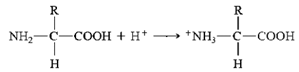
• Proteins are composed of amino acids
• Contain at least one carboxyl group (COOH) and at least one amino group (NH2)
• When pH is high - Free carboxyl group at one end of a protein acts like an acid and releases H+

• When pH is low - Free amino group at the other end of a protein can act as a base by combining with H+
Hemoglobin as buffer in red blood cells
• Carbon dioxide (CO2) passes from tissue cells into red blood cells
• Combines with water (H2O) to form carbonic acid (H2CO3)

• Oxyhemoglobin (Hb O2) is giving up its oxygen to tissue cells
• Reduced Hb (deoxyhemoglobin) picks up most of the H+

b. Carbonic Acid–Bicarbonate Buffer System
• The bicarbonate ion (HCO3), act as a weak base
• Carbonic acid (H2CO3), act as a weak acid
When excess of H+ - HCO3 - can function as a weak base and remove the excess H+

When shortage of H+ - H2CO3 can function as a weak acid and provide H+

c. Phosphate Buffer System
Components - Ions Dihydrogen phosphate (H2PO4-) and Monohydrogen phosphate (HPO42-) when pH is high
• Dihydrogen phosphate ion acts as a weak acid and buffer strong bases such as OH-
When pH is low
• Monohydrogen phosphate ion buffers the H+ released by a strong acid & acts as a weak base
II. Exhalation of carbon dioxide
• Increase in the carbon dioxide (CO2) concentration in body fluids increases H+ concentration and thus lowers the pH
• H2CO3 can be eliminated by exhaling CO2, it is called a volatile acid
• A decrease in the CO2 concentration of body fluids raises the pH
III. Kidney excretion of H+
• In PCT, Na+/H+ antiporters secrete H+ as they reabsorb Na+
• In PCT, Na+/H+ antiporters secrete H+ as they reabsorb
• In collecting ducts, intercalated cells absorb K+ & HCO3; secrete H+

Acid–Base Imbalances
Normal pH range of systemic arterial blood is between 7.35 – 7.45
Acidosis (or acidemia)
• Condition where blood pH is below 7.35
• Results in depression of the CNS
Alkalosis
• Conditions where blood pH is above 7.45
• Results in over excitability of the CNS

Clearance tests
• Measures GFR; useful in assessing renal function
• Volume of plasma that would be completely cleared of a substance per min
• Clearance (C), expressed as ml/min, can be calculated by using the formula
C = U х V / P
Where, U = Concentration of substance in urine
V = Volume of urine in ml excreted per min
P = Concentration of substance in plasma
• Measurement of clearance of substance already present in the blood is preferred
• The compounds measured are: Creatinine and urea
Creatinine clearance test
• Creatinine is the excretory product derived from creatinine phosphate
• Its excretion is constant and not influenced by body metabolism and dietary factor
• It is the volume of plasma that would be completely cleared of creatinine per min
C =U х V / P
Where, U = Concentration of creatinine in urine
V = Volume of urine in ml excreted per min
P = Concentration of creatinine in plasma
Importance
• Values are close to GFR, hence measurement is sensitive to assess the renal glomerular filtration
• Decrease in clearance value serves as an indicator of decreased GFR due to renal damage
Urea clearance test
• Urea is the end product of protein metabolism
• It is the volume (ml) of plasma that would be completely cleared of urea per min
Cm = U х V / P
Where, Cm = Maximum urea clearence (only when urine output is > 2 ml/ min
U = Concentration of urea in urine, V = Volume of urine in ml excreted per min
P = Concentration of urea in plasma
• When urine output is less than 2 ml/min, it is called standard urea clearence
Cs = U х (V) 1/2
P
• Urea clearence is less than GFR
• After being filtered by glomeruli, it is partially reabsorbed by the renal tubule
• Influenced by protein content of diet
Importance
• Value below 75%, indicator of renal damage
• If clearance is below 50%, blood urea levels increases
Urine transportation, storage and elimination
• Urine from collecting duct transported through minor calyx, major calyx, renal pelvis and later drained into urinary bladder through ureters
• Urine is then discharged from the body through the single urethra

Ureters
• Two in number, Retroperitoneal
• 25-30 cm in length; Diameter varies from 1mm to 10mm
• Curved medially & pass obliquely through posterior aspect of urinary bladder
• Carry urine from renal pelvis to urinary bladder
• Peristaltic contraction, hydrostatic pressure & gravity are the contributing force for urine flow
Layers of ureter
3 layers of tissue that form the wall of ureter
– Mucosa; Musclaris; Aventitia
Mucosa
• Mucous membrane with transitional epithelium
• Able to stretch; accommodate variable amount of urine
• Goblet cells produce mucus, prevents cells from coming in contact with urine
Musclaris
• Intermediate coat
• Has inner longitudinal; outer circular smooth muscle fibres
• In the distal third, ureter is made of inner longitudinal, middle circular, outer longitudinal
• Function – Peristalsis
Adventitia
• Superficial coat
• Aerolar connective tissue containing blood vessels, lymphatic vessels & nerves
• Blends in with the surrounding tissue
• Anchors ureter in place
Urinary Bladder
• Hollow, muscular organ
• Situated in the pelvic cavity, posterior to pubic symphysis
• Peritoneum holds the urinary bladder in place
• Usually pear shaped
• Capacity averages 700 - 800 ml
Anatomy and histology of urinary bladder
• In the floor of urinary bladder is a small triangular area, trigone
• Two posterior corners of trigone contain two urethral opening & one external urethral opening, the internal urethral orifice

Layers of urinary bladder
Walls of urinary bladder, made up of 3 coats
Mucosa
• Mucus membrane with transitional epithelium
• Rugae, permits expansion of urinary bladder
Muscularis/ Detrusor muscle
• Has 3 layers of smooth muscle fibres - Inner longitudinal; Middle circular; Outer longitudinal
• External urethral sphincter is made of skeletal muscle
Adventitia
• Layer of aerolar connective tissue
• Continuous with that of uterus
• Over the superior surface of urinary bladder is serosa, a layer of visceral peritoneum
Micturition Reflex
• Discharge of urine from urinary bladder is micturition or urination or Voiding
Volume of urine in urinary bladder exceeds 200- 400 ml
â
Pressure in bladder increases
â
Stretch receptors transmit nerve impulses to spinal cord
â
Impulses reaches micturition centre in S2, S3
â
Trigger spinal reflex, Micturition reflex
• Micturition reflex discharge urine from urinary bladder via parasympathetic impulses
• Contraction of detrusor muscle occur
• Relaxation of internal urethral sphincter muscle
Urethra
• Small tube leading from internal urethral orifice of urinary bladder to exterior of the body
• Helps to discharge urine from the body
• Anatomy and histology of urethra differ in females and males
In female,
• Urethra lie posterior to pubic symphysis
• External urethral orifice located between clitoris and vagina
• Wall consists of deep mucosa and muscularis
In males,
• Urethra extends from internal urethral orifice to the exterior
• First passes through prostate, then through perineum & finally through penis
• Wall of male urethra consists of deep mucosa and superfacial muscularis subdivide into
a. Prostatic urethra – passes through prostate
b. Membranous urethra – passes through perineum
c. Spongy urethra – longest portion, passes through penis
Diseases of the urinary system
Renal calculi
• Crystals of salts present in urine occasionally precipitate and solidify into insoluble stones
• Contains crystals of calcium oxalate, uric acid & calcium phosphate
Causes -Ingestion of excessive calcium
- Low water intake
- Abnormally alkaline or acidic urine
- Over activity of parathyroid glands
Urinary tract infection
• Describes either an infection of a part of urinary system or presence of large number of microbes in urine
• More common in female due to shorter length of urethra
• Includes urethritis, cystitis, pyelonephritis
Symptoms –Painful or burning urination
– Frequent urination
– Low back pain
– Bed wetting
Glomerular diseases
Glomerulo nephritis
• Inflammation of kidney involving glomeruli
• Due to allergic reaction to the toxins produced by streptococcal bacteria
• Symptoms – Haematuria, proteinuria
Nephrotic syndromes
• Condition characterised by proteinuria, hyperlipidemia
• Causes- DM, SLE, Cancer & AIDS
• Symptoms- Edema around eyes, ankle, feet and abdomen
Renal failure
Decrease or cessation of glomerular filteration
Acute renal failure
• Kidneys abruptly stops working entirely
• Causes – Low blood volume, decreased cardiac output, damaged renal tubules, kidney stones, NSAIDS
• Symptoms – Oliguria, Anuria, Anaemia
Chronic renal failure
• Progressive and usually irreversible decline in GFR
• Causes – chronic glomerulonephritis, pyelonephritis, Polycystic kidney disorders, Traumatic loss of kidney tissues
Polycystic kidney disease (PKD)
• Inherited disorder, kidneys get riddled with 100 – 1000 of cysts (fluid filled cavities)
• Progressive impairment of renal function leading to end stage renal failure
Summary
• The organs of the urinary system are the kidneys, ureters, urinary bladder, and urethra
• Kidneys filter blood and return most water and many solutes to the bloodstream, the remaining water and solutes constitute urine
• Kidneys are retroperitoneal organs; three layers of kidney – renal capsule, adipose capsule, and renal fascia
• Nephron is the functional unit of the kidneys
• Nephron consists of a renal corpuscle and a renal tubule
• Nephrons perform three basic tasks: glomerular filtration, tubular secretion, and tubular reabsorption
• Renal autoregulation, neural regulation, and hormonal regulation determines the renal function
• Renin angiotensin and aldosterone system helps in maintaining the blood volume by releasing renin, activating angiotensin II, and releasing aldosterone
• Various mechanisms help maintain the pH of systemic arterial blood between 7.35 and 7.45
• Buffers act to temporarily bind H+, removing the highly reactive, excess H+ from solution
• Increase in concentration of CO2 lowers the pH and will be reduced by exhalation of CO2
• Kidney excretes excess of H+ ions to maintain the pH of blood
• Acid- base imbalance causes acidosis and alkalosis
• Various clearance tests helps in measuring the GFR and are useful in assessing renal function
• Ureters are retroperitoneal and consist of a mucosa, muscularis, and adventitia
• They transport urine from the renal pelvis to the urinary bladder, primarily via peristalsis
• Urinary bladder is located in the pelvic cavity; its function is to store urine before micturition
• Urethra is a tube leading from the floor of the urinary bladder to the exterior
• Discharge of urine from urinary bladder is micturition
• Micturition reflex discharges urine from the urinary bladder via parasympathetic impulses
• Diseases associated with urinary system include renal calculi, urinary tract infection, glomerular disease, renal failure and polycystic kidney disease
0 Comments: