

Cancer - B. Pharma 2nd Semester Pathophysiology notes pdf
Cancer
Content
Cancer
• Classification
• Spread of cancer
Objectives
At the end of this lecture, student will be able to
• Define the term “Tumor”
• List main characteristics of benign and malignant cancer
• Describe the nomenclature used for various types of tumors
• Explain the mechanism of spread of tumors
CANCER
General biology of cancer
Neoplasm/ Tumour - “A mass of tissue formed as a result of abnormal, excessive, uncoordinated, autonomous and purposeless proliferation of cells”
• Oncology– Branch of science dealing with the study of neoplasm
• Transformation from a living normal cell into a living tumor cell
Basic features of change in neoplasia
• Change is irreversible; becomes fixed character of a transformed cell
• Acquired fixed character is heritable; tumor cell divide to give tumor cell
• Change once occurred is self-perpetuating
• Tumor cell has uncontrolled passion for continued proliferation
Classification of tumors
• Based on the nature of tumors, they are classified as
Benign tumor – Harmless and self-limited
Malignant tumor – Harmful and rapidly growing
• Names of every tumor ends with ‘oma’
• Malignant tumor of epithelial tissue – Carcinoma
• Malignant tumor of connective tissue - Sarcoma
Classification of tumor based on tissue of origin
| Tissue of origin | Benign | Malignant |
| Epithelial tumors | ||
| 1. Squamous epithelium | Squamous cell papilloma | Squamous cell carcinoma |
| 2. Transitional epithelium | Transitional epithelium papilloma | Transitional epithelium carcinoma |
| 3. Glandular epithelium | Adenoma | Adenocarcinoma |
| 4. Hepatocytes | Liver cell adenoma | Hepatocellular carcinoma (Hepatoma) |
| Tissue of origin (Non epithelial tumors) | Benign | Malignant |
| Adipose tissue | Lipoma | Liposarcoma |
| Fibrous tissue | Fibroma | Fibrosarcoma |
| Cartilage | Chondroma | Chondrosarcoma |
| Bone | Osteoma | Osteosarcoma |
| Blood vessels | Haemangioma | Angiosarcoma |
| Nerve cells | Ganglia Neuroma | Neuroblastoma |
Contrasting features of benign and malignant tumor
| Features | Benign | Malignant |
| Macroscopic features | ||
| Encapsulated/ well circumscribed | Irregular & poorly circumscribed |
| 2. Surrounding tissues | Often compressed | Usually invaded |
| Usually small | Often large |
| 4. Secondary changes | Occurs less often | Occurs more often |
| Microscopic Features | ||
| Closely resembles the tissue of origin | Poor resemblance to the tissue of origin |
| 2. Basal polarity | Retained | Lost |
| 3. Pleomorphism | Normal | Increased |
| Features | Benign | Malignant |
| Microscopic features (cont..d) | ||
| 4. Neuclio- cytoplasmic ratio | Normal | Increased |
| Absent | Present |
| Always typical mitosis | Atypical & abnormal mitosis |
| 7. Tumor giant cells | May be present but with atypical nucleus | Always present with atypical nucleus |
| 8. Cytoplasm | With normal constituents | Elements are reduced or lost |
| 9. Functions | Usually well maintained | Retained/ lost/ abnormal |
| Growth rate | Usually slow | Rapid |
| Local invasion | Often compresses the surroundings; no invasion/ infiltration | Invade & infiltrate the adjacent tissue |
| Metastatis (Spreading) | Absent | Present |
Structure of tumor
Tumor mass consists of:
- Parenchyma
- Stroma
Parenchyma
• Formed by proliferating tumor cells
• Parenchyma of benign tumor – organised pattern with resemblance to tissue of origin, differentiation
• Parenchyma of malignant tumor – unorganised, atypical, distorted, relation of tumor cell with basement is lost, anaplasia
Stroma
• Supporting tissue of tumor
• Consists of fibrous tissue carrying blood vessels for nourishing tumor cell
• More malignant the tumor, Cirrhous
• Carcinoma with scanty stroma – celluloid & medullary
• New blood vessels form from pre existing onesless is the fibrous tissue
• Carcinoma with extensive stroma – with the help of a factor, “tumor angiogenesis factor”
Spread of cancer
(Two mechanism for the spread of cancer)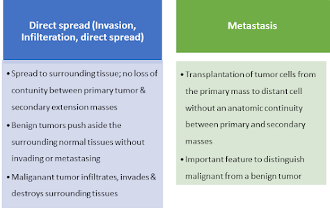
Routes of spread of cancer
1. Infiltration of tissue spaces
• Tissue spaces - preformed passages; paths of least resistance
• Most vulnerable tissues – soft tissues – adipose, muscle,
• Gamete/ compact tissues like capsule of organs, cartilage and bone (not marrow) offer greater resistance
• Tissue subjected to infiltration are destroyed by the proteolytic enzymes & lytic substances elaborated by cancer tissue
• Tissue space invasion brings the tumor cell in direct contact with normal cells, lymphatic and blood vessels
2. Hematogenous spread:
• Carcinoma of lungs, thyroid, kidney and the prostate spread through blood vessels
Tumor cells enter blood stream by 2 ways
- Via thorasic duct – either by perforation of vein or by lymphatic drainage
- By direct invasion of blood vessels (large veins, venules & capillaries); arteries not involved due to their thick wall
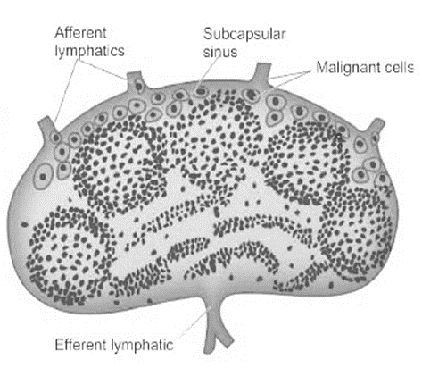
3. Spread via lymphatics: Most common with carcinoma; results in both invasion & metastasis

Spread via lymphatics
Lymphatic spread begins by lodgement of tumour cells in subcapsular sinus via afferent lymphatics entering at the convex surface of the lymph node
Hematogenous spread

4. Spread via serous sacs
• Spread through peritoneal cavity; common in cancer of GIT & ovary
• Trans pleural spread - in carcinoma of lungs and breast
• Trans pericardial spread may also occur
5. Spread along epithelium line surfaces
• Intact epithelium, mucous coat acquires resistance for penetration of tumor
• Implantation tumor – tumor spread along the surface of epithelium
6. Spread via CSF
• Cerebrospinal cavities are affected by the escape of tumor cells from
• the malignant tumor in the brain or meninges
Summary
• A tumour is a mass of tissue formed as a result of abnormal, excessive, uncoordinated, autonomous and purposeless proliferation of cells
• Tumors are classified as benign and malignant
• Benign tumors are harmless and do not spread while malignant tumors are harmful and spread
• Tumor is made up of parenchyma and stroma
• Tumor spread by two mechanism – Haematogenous spread and lymphatic spread

0 Comments: